The doctoral dissertation in the field of Medicine will be examined at the Faculty of Health Sciences.
What is the topic of your doctoral research? Why is it important to study the topic?
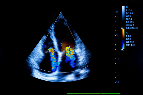
My doctoral research focuses on advanced imaging techniques of coronary arteries, myocardial function, and ischemia. Coronary artery disease remains a significant clinical problem globally, and successful therapy development is based on accurate detection by various diagnostic methods. Noninvasive imaging techniques have improved the evaluation of patients with known or suspected coronary artery disease. Thus, advanced imaging techniques provide important diagnostic and prognostic information for managing coronary artery disease.
What are the key findings or observations of your doctoral research?
My doctoral thesis is an article-based dissertation and consists of five sub-studies. In general, the sub-studies were different in aims, setting, and designs. However, the results of these studies provide additional information on the applicability and clinical utility of non-invasive imaging modalities and encourage a more widespread utilization of these modalities. The results support the concept that echocardiography allows accurate and comprehensive evaluation of cardiac systolic function in coronary artery disease including acutely ill patients. The meta-analysis provides novel information on the diagnostic performance of non-invasive diagnostic tests in evaluation of suspected chronic coronary artery disease depending on clinical likelihood of disease. Anatomical imaging, such as computed tomography angiography, accurately rules out obstructive disease. Functional imaging, such as myocardial perfusion imaging, is the most accurate for ruling in obstructive disease.
How can the results of your doctoral research be utilised in practice?
In the first sub-study, the area-length methods obtained could be the valid alternative options for assessing the left ventricle ( LV) volume, in some difficult situations like in intensive care unit ( ICU). In the second sub-study, the short-axis echocardiography strains could be complementary predictors for LV remodeling and markers of residual viable myocardium in patients with Myocardial infarction (MI). The third sub-study results suggest that the new drug (Xenon) could protect against myocardial dysfunction after cardiac arrest and in addition, the strain echocardiography is a new promising method and could improve the diagnostic and prognostic accuracy of echocardiography in the ICU clinical setting. The fourth sub-study results recommended using the strain parameters to improve the accuracy of echocardiography for detection of reversible myocardial dysfunction. The results of the fifth substudy can be directly implemented as an aid to select most appropriate diagnostic test for patients with suspected obstructive coronary artery disease depending on clinical likelihood of disease. The clinical value was highlighted by inclusion of the study results in the 2019 European Society of Cardiology (ESC) Guidelines for diagnosing and managing chronic coronary syndromes.
What are the key research methods and materials used in your doctoral research?
My doctoral study consists of five sub-studies. The first and second Sub-studies were conducted on an experimental model of chronic coronary artery disease. The purpose of the sub-study I was to validate echocardiographic area-length methods. Sub-study II was performed to assess the accuracy of regional short-axis myocardial strain in detecting chronic MI and to evaluate the relations between myocardial strain values and the heart failure process. The third sub-study was a clinical trial performed to evaluate a new drug (Xenon). The echocardiography was performed to assess the effect of xenon on left ventricular function in comatose survivors of out-hospital cardiac arrest from a cardiac cause. The fourth and fifth Sub-studies were systematic reviews and meta-analyses. The fourth Sub-study was a meta-analysis for all prospective trials assessing the reversible myocardial dysfunction after revascularization using a new method (Speckle tracking echocardiography). The fifth Sub-studies was a meta-analysis performed to evaluate the diagnostic performance of different modalities in detecting anatomically and functionally significant coronary artery disease.
This doctoral study was carried out in collaboration between the University of Eastern Finland and the University of Turku. The clinical and experimental parts of my doctoral study were performed mainly in departments and labs of Turku University Hospital. I am very grateful to the whole research group in Turku and Kuopio for their efforts during my doctoral study.
The doctoral dissertation of Haitham Ballo, MBChB, entitled Advanced cardiac imaging of cardiac function and coronary artery disease will be examined at the Faculty of Health Sciences. The doctoral defence will take place at Turku University Hospital and it will be streamed online. The Opponent in the public examination will be Professor Katriina Aalto-Setälä of Tampere University, and the Custos will be Professor Antti Saraste of the University of Turku. The event will be held in English.
Doctoral defence online
For further information, please contact:
Haitham Ballo, hballo (a) uef.fi, tel. 0466406734


