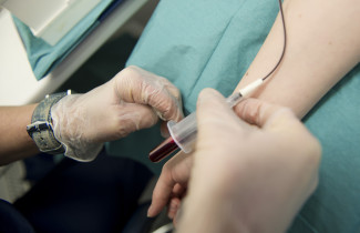
The neurofilament light chain protein (NfL) aids the diagnostic process of frontotemporal lobar degeneration (FTLD). It correlates with the disease prognosis and the brain atrophy rate recognized by automatic MRI quantifying methods, shows the PhD thesis made by MD Antti Cajanus at the University of Eastern Finland.
Frontotemporal lobar degeneration (FTLD) is the second most common early-onset neurodegenerative disorder after Alzheimer’s disease (AD). It is characterised by a change in a patient’s behavior and personality or with problems with speech and language. Several genetic etiologies have been recognized to cause a predisposition towards developing FTLD, yet the heterogeneity of the clinical phenotype hinders the diagnostic process. Furthermore, current clinical work is lacking sensitive and specific biomarkers. As a consequence, delays in the diagnosis, and misdiagnosis often occurs, leading to ineffective and potentially harmful treatments, increased healthcare costs, and patient and caregiver distress.
The thesis aimed to evaluate MRI biomarkers and blood-derived NfL for detecting early phase FTLD. NfL was found to be an excellent tool for differentiating between FTLD and primary psychiatric disorders (PPD). Furthermore, patients with higher serum NfL levels had significantly shorter survival times and faster rates of brain atrophy, indicative of a strong correlation with the disease severity and prognosis. The results showed that automated structural MRI quantification methods were able to distinguish FTLD from other neurodegenerative disorders with similar sensitivity to an experienced neuroradiologist. Also, the severity of behavioral symptoms of the disease were found to correlate with the volume of the frontal lobe cortex.
“To date, there is no curative nor disease-modifying treatment for slowing its progression. Given that specific underlying pathologies require specific pharmaceutical treatments, it is essential to distinguish the pathology in vivo for an adequate selection of study subjects. Furthermore, future clinical trials aimed at treatment need biomarkers for assessing disease severity, prognosis, and positive therapeutic effects.”
The doctoral dissertation of MD Antti Cajanus entitled ”Diagnostic Brain Imaging and Blood Biomarkers for Early Frontotemporal Lobar Degeneration” will be examined at the Faculty of Health Sciences, University of Eastern Finland. The Opponent in the public examination will be Professor Juha Rinne of the University of Turku, and the Custos will be Professor Anne Remes of the University of Oulu. The public examination will be held in Finnish on 11th December 2020 at 12 noon and it can be followed online.
Photo available for download at https://mediabank.uef.fi/A/UEF+Media+Bank/38073?encoding=UTF-8


